9I53 | pdb_00009i53

Crystal structure of the SARS-CoV-2 helicase NSP13 in complex with ATP


Experimental Data Snapshot

  • Method: X-RAY DIFFRACTION
  • Resolution: 1.92 Å
  • R-Value Free: 
    0.229 (Depositor), 0.229 (DCC) 
  • R-Value Work: 
    0.187 (Depositor), 0.187 (DCC) 
  • R-Value Observed: 
    0.189 (Depositor) 

Starting Model: experimental
View more details

wwPDB Validation   3D Report Full Report


Ligand Structure Quality Assessment 


This is version 1.0 of the entry. See complete history


Literature

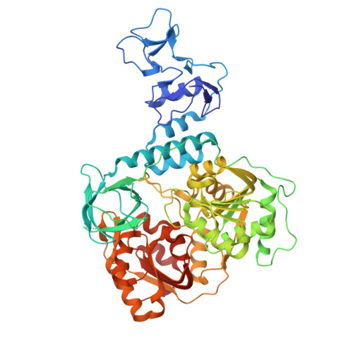
Nucleotide-bound crystal structures of the SARS-CoV-2 helicase NSP13.

Kloskowski, P.Neumann, P.Berndt, A.Ficner, R.

(2025) Acta Crystallogr F Struct Biol Commun 

  • DOI: https://doi.org/10.1107/S2053230X25005266
  • Primary Citation of Related Structures:  
    9I53

  • PubMed Abstract: 

    Nucleotide-bound crystal structures of SARS-CoV-2 NSP13 in ADP- and ATP-bound states were resolved to 1.8 and 1.9 Å, respectively. The ADP-bound model captures a state immediately following ATP hydrolysis, with both ADP and orthophosphate still present in the active site. Further comparative analysis revealed that crystal packing influences NSP13 by stabilizing the nucleotide-binding site, underscoring the importance of accounting for these effects in structure-based drug design targeting NSP13.


  • Organizational Affiliation

    Department of Molecular Structural Biology, Institute of Microbiology and Genetics, Göttingen Center of Molecular Biosciences (GZMB), University of Göttingen, Justus-von-Liebig-Weg 11, 37077 Göttingen, Germany.


Macromolecules
Find similar proteins by:  (by identity cutoff)  |  3D Structure
Entity ID: 1
MoleculeChains Sequence LengthOrganismDetailsImage
SARS-CoV-2 helicase NSP13
A, B
603Severe acute respiratory syndrome coronavirus 2Mutation(s): 0 
Gene Names: rep1a-1b
EC: 3.4.19.12 (PDB Primary Data), 3.4.22 (PDB Primary Data), 3.4.22.69 (PDB Primary Data), 2.7.7.48 (PDB Primary Data), 3.6.4.12 (PDB Primary Data), 3.6.4.13 (PDB Primary Data), 3.1.13 (PDB Primary Data), 3.1 (PDB Primary Data), 2.1.1 (PDB Primary Data)
UniProt
Find proteins for P0DTD1 (Severe acute respiratory syndrome coronavirus 2)
Explore P0DTD1 
Go to UniProtKB:  P0DTD1
Entity Groups  
Sequence Clusters30% Identity50% Identity70% Identity90% Identity95% Identity100% Identity
UniProt GroupP0DTD1
Sequence Annotations
Expand
  • Reference Sequence
Small Molecules
Ligands 4 Unique
IDChains Name / Formula / InChI Key2D Diagram3D Interactions
ATP (Subject of Investigation/LOI)
Query on ATP

Download Ideal Coordinates CCD File 
F [auth A],
I [auth A],
M [auth B]
ADENOSINE-5'-TRIPHOSPHATE
C10 H16 N5 O13 P3
ZKHQWZAMYRWXGA-KQYNXXCUSA-N
MPO
Query on MPO

Download Ideal Coordinates CCD File 
H [auth A],
O [auth B]
3[N-MORPHOLINO]PROPANE SULFONIC ACID
C7 H15 N O4 S
DVLFYONBTKHTER-UHFFFAOYSA-N
PO4
Query on PO4

Download Ideal Coordinates CCD File 
G [auth A],
N [auth B],
P [auth B]
PHOSPHATE ION
O4 P
NBIIXXVUZAFLBC-UHFFFAOYSA-K
ZN
Query on ZN

Download Ideal Coordinates CCD File 
C [auth A]
D [auth A]
E [auth A]
J [auth B]
K [auth B]
C [auth A],
D [auth A],
E [auth A],
J [auth B],
K [auth B],
L [auth B]
ZINC ION
Zn
PTFCDOFLOPIGGS-UHFFFAOYSA-N
Experimental Data & Validation

Experimental Data

  • Method: X-RAY DIFFRACTION
  • Resolution: 1.92 Å
  • R-Value Free:  0.229 (Depositor), 0.229 (DCC) 
  • R-Value Work:  0.187 (Depositor), 0.187 (DCC) 
  • R-Value Observed: 0.189 (Depositor) 
Space Group: P 1
Unit Cell:
Length ( Å )Angle ( ˚ )
a = 59.577α = 102.99
b = 70.643β = 95.29
c = 85.885γ = 112.53
Software Package:
Software NamePurpose
PHENIXrefinement
autoPROCdata reduction
autoPROCdata scaling
REFMACphasing

Structure Validation

View Full Validation Report


Ligand Structure Quality Assessment 


Entry History & Funding Information

Deposition Data


Funding OrganizationLocationGrant Number
German Research Foundation (DFG)Germany--

Revision History  (Full details and data files)

  • Version 1.0: 2025-07-23
    Type: Initial release