9OMS | pdb_00009oms

X-ray structure of human acetylcholinesterase (hAChE) in complex with bis-oxime reactivator LG-1922


Experimental Data Snapshot

  • Method: X-RAY DIFFRACTION
  • Resolution: 2.40 Å
  • R-Value Free: 
    0.219 (Depositor), 0.218 (DCC) 
  • R-Value Work: 
    0.192 (Depositor), 0.193 (DCC) 
  • R-Value Observed: 
    0.194 (Depositor) 

Starting Model: experimental
View more details

wwPDB Validation   3D Report Full Report


This is version 1.0 of the entry. See complete history


Literature

Kinetic and structural evidence for specific DMSO interference with reversible binding of uncharged bis-oximes to hAChE and their reactivation kinetics of OP-hAChE.

Kolic, D.Gerlits, O.Kucharski, M.Gorecki, L.Joiner, N.Kovalevsky, A.Radic, Z.

(2025) Chem Biol Interact 419: 111649-111649

  • DOI: https://doi.org/10.1016/j.cbi.2025.111649
  • Primary Citation of Related Structures:  
    9OMS, 9OMT

  • PubMed Abstract: 

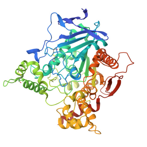
    The structural basis of inhibitory effect of organic solvent dimethyl sulfoxide (DMSO) on human acetylcholinesterase (EC 3.1.1.7; hAChE) was inferred from the effect of DMSO on kinetics of reversible inhibition of uncharged, heterocyclic bis-oximes to hAChE, from DMSO effect on rates of reactivation of inactive organophosphate (OP)-hAChE conjugates by bis-oximes and by X-ray structures of bis-oxime and DMSO binding to hAChE. The reversible inhibition constant of DMSO for hAChE in 0.1 M phosphate buffer pH 7.4 at 22 °C, was K i = (0.32 ± 0.04) % (or 45 ± 5 mM). The K i of the bis-oxime LG-703 for hAChE was 3.2-fold larger in 1 % DMSO, consistent with direct competition between LG-703 and DMSO. The X-ray structure of the LG-703∗hAChE complex (PDB ID: 6U3P) shows DMSO and LG-703 bound to individual hAChE monomers, LG-703 in the chain A and DMSO in the chain B. In the co-crystallization both small molecules were present at a similar excess over their corresponding K i values for hAChE (7.8-fold for DMSO and 6.5-fold for LG-703) and formation of two different complexes (DMSO∗hAChE and LG-703∗hAChE), in the same crystal, appears consistent with inhibition kinetics. Furthermore, rates of reactivation of paraoxon-inhibited hAChE (POX-hAChE) and of VX-hAChE by LG-703 and by a novel heterocyclic bis-oxime LG-1922 were reduced 2 - 3-fold in DMSO, consistent with observation of the active-center-bound DMSO molecules in the newly solved structure of the LG-1922∗POX-hAChE complex presented here and in our POX-hAChE structure (PDB ID: 8DT2) showing obstruction of the reactivator access to the conjugated P atom.


  • Organizational Affiliation

    Institute for Medical Research and Occupational Health, HR-10001, Zagreb, Croatia.


Macromolecules
Find similar proteins by:  (by identity cutoff)  |  3D Structure
Entity ID: 1
MoleculeChains Sequence LengthOrganismDetailsImage
Acetylcholinesterase
A, B
550Homo sapiensMutation(s): 0 
Gene Names: ACHE
EC: 3.1.1.7
UniProt & NIH Common Fund Data Resources
Find proteins for P22303 (Homo sapiens)
Explore P22303 
Go to UniProtKB:  P22303
PHAROS:  P22303
GTEx:  ENSG00000087085 
Entity Groups  
Sequence Clusters30% Identity50% Identity70% Identity90% Identity95% Identity100% Identity
UniProt GroupP22303
Sequence Annotations
Expand
  • Reference Sequence
Small Molecules
Ligands 3 Unique
IDChains Name / Formula / InChI Key2D Diagram3D Interactions
A1CD5 (Subject of Investigation/LOI)
Query on A1CD5

Download Ideal Coordinates CCD File 
C [auth A],
G [auth B]
2-(hydroxyimino)-N-{2-[(3S)-1-(3-{[(2E)-2-(hydroxyimino)acetyl]amino}propyl)piperidin-3-yl]ethyl}acetamide
C14 H25 N5 O4
KZXNYUVBCIGYGX-XZXJBNDFSA-N
GOL
Query on GOL

Download Ideal Coordinates CCD File 
E [auth A],
F [auth A],
H [auth B],
I [auth B]
GLYCEROL
C3 H8 O3
PEDCQBHIVMGVHV-UHFFFAOYSA-N
NO3
Query on NO3

Download Ideal Coordinates CCD File 
D [auth A]NITRATE ION
N O3
NHNBFGGVMKEFGY-UHFFFAOYSA-N
Experimental Data & Validation

Experimental Data

  • Method: X-RAY DIFFRACTION
  • Resolution: 2.40 Å
  • R-Value Free:  0.219 (Depositor), 0.218 (DCC) 
  • R-Value Work:  0.192 (Depositor), 0.193 (DCC) 
  • R-Value Observed: 0.194 (Depositor) 
Space Group: P 31
Unit Cell:
Length ( Å )Angle ( ˚ )
a = 124.47α = 90
b = 124.47β = 90
c = 129.9γ = 120
Software Package:
Software NamePurpose
PHENIXrefinement
HKL-3000data scaling
HKL-3000data reduction
PHASERphasing

Structure Validation

View Full Validation Report


Entry History & Funding Information

Deposition Data


Funding OrganizationLocationGrant Number
National Institutes of Health/National Institute of Neurological Disorders and Stroke (NIH/NINDS)United StatesR21NS120839

Revision History  (Full details and data files)

  • Version 1.0: 2025-07-30
    Type: Initial release