9KUD | pdb_00009kud

Crystal structure of SARS-CoV-2 JN.1 variant RBD complexed with squirrel ACE2


Experimental Data Snapshot

  • Method: X-RAY DIFFRACTION
  • Resolution: 3.14 Å
  • R-Value Free: 
    0.281 (Depositor), 0.268 (DCC) 
  • R-Value Work: 
    0.225 (Depositor), 0.228 (DCC) 
  • R-Value Observed: 
    0.227 (Depositor) 

Starting Model: experimental
View more details

wwPDB Validation   3D Report Full Report


Ligand Structure Quality Assessment 


This is version 1.1 of the entry. See complete history


Literature

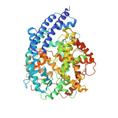
Cross-species recognition of squirrel ACE2 by the receptor binding domains of SARS-CoV-2, RaTG13, PCoV-GD and PCoV-GX.

Wang, C.Nan, X.Deng, Y.Fan, S.Lan, J.

(2025) Structure 

  • DOI: https://doi.org/10.1016/j.str.2025.07.003
  • Primary Citation of Related Structures:  
    9JR4, 9JR5, 9JR7, 9JRC, 9KUD

  • PubMed Abstract: 

    SARS-CoV-2 has a broad animal host range, but its origin and intermediate hosts are still debated. Arctic ground squirrel is proved to be a susceptible animal host of SARS-CoV-2 and RaTG13, as the Arctic ground squirrel ACE2 (sACE2) could bind to the RBDs of SARS-CoV-2 and RaTG13, and support transduction of the SARS-CoV-2 and RaTG13 pseudovirions. Here, we determined crystal structures of sACE2 bound to the RBDs of RaTG13, PCoV-GD, PCoV-GX, SARS-CoV-2 A372T and SARS-CoV-2 JN.1 variant. SPR assay indicated that RBD residues 493, 498 and 501 might be important for sACE2 recognition. Notably, N322-linked glycans of sACE2 were found to be in contact with S375 of the RaTG13 RBD. Moreover, the recent SARS-CoV-2 KP.2 and KP.3 variant RBDs could also bind to the sACE2. Our results provide further insights into interactions between coronavirus RBD and cell receptor ACE2, which may promote our understanding of SARS-CoV-2 evolution.


  • Organizational Affiliation

    School of Biomedical Sciences, Hunan University, Changsha, Hunan, China.


Macromolecules
Find similar proteins by:  (by identity cutoff)  |  3D Structure
Entity ID: 1
MoleculeChains Sequence LengthOrganismDetailsImage
Angiotensin-converting enzyme
A, B
596Petaurus norfolcensisMutation(s): 0 
Gene Names: ACE2
EC: 3.4
UniProt
Find proteins for A0A8D2KIZ1 (Urocitellus parryii)
Explore A0A8D2KIZ1 
Go to UniProtKB:  A0A8D2KIZ1
Entity Groups  
Sequence Clusters30% Identity50% Identity70% Identity90% Identity95% Identity100% Identity
UniProt GroupA0A8D2KIZ1
Glycosylation
Glycosylation Sites: 3
Sequence Annotations
Expand
  • Reference Sequence
Find similar proteins by:  (by identity cutoff)  |  3D Structure
Entity ID: 2
MoleculeChains Sequence LengthOrganismDetailsImage
Spike protein S1C [auth E],
D [auth F]
197Severe acute respiratory syndrome coronavirus 2Mutation(s): 18 
Gene Names: S2
UniProt
Find proteins for P0DTC2 (Severe acute respiratory syndrome coronavirus 2)
Explore P0DTC2 
Go to UniProtKB:  P0DTC2
Entity Groups  
Sequence Clusters30% Identity50% Identity70% Identity90% Identity95% Identity100% Identity
UniProt GroupP0DTC2
Glycosylation
Glycosylation Sites: 2Go to GlyGen: P0DTC2-1
Sequence Annotations
Expand
  • Reference Sequence
Oligosaccharides

Help

Entity ID: 3
MoleculeChains Length2D Diagram Glycosylation3D Interactions
alpha-D-mannopyranose-(1-6)-beta-D-mannopyranose-(1-4)-2-acetamido-2-deoxy-beta-D-glucopyranose-(1-4)-2-acetamido-2-deoxy-beta-D-glucopyranoseE [auth C]4N-Glycosylation
Small Molecules
Ligands 2 Unique
IDChains Name / Formula / InChI Key2D Diagram3D Interactions
NAG
Query on NAG

Download Ideal Coordinates CCD File 
F [auth A]
G [auth A]
H [auth A]
K [auth E]
L [auth E]
F [auth A],
G [auth A],
H [auth A],
K [auth E],
L [auth E],
M [auth F]
2-acetamido-2-deoxy-beta-D-glucopyranose
C8 H15 N O6
OVRNDRQMDRJTHS-FMDGEEDCSA-N
ZN
Query on ZN

Download Ideal Coordinates CCD File 
I [auth A],
J [auth B]
ZINC ION
Zn
PTFCDOFLOPIGGS-UHFFFAOYSA-N
Experimental Data & Validation

Experimental Data

  • Method: X-RAY DIFFRACTION
  • Resolution: 3.14 Å
  • R-Value Free:  0.281 (Depositor), 0.268 (DCC) 
  • R-Value Work:  0.225 (Depositor), 0.228 (DCC) 
  • R-Value Observed: 0.227 (Depositor) 
Space Group: P 21 21 21
Unit Cell:
Length ( Å )Angle ( ˚ )
a = 128.674α = 90
b = 129.157β = 90
c = 138.062γ = 90
Software Package:
Software NamePurpose
PHENIXrefinement
HKL-2000data reduction
HKL-2000data scaling
PHENIXphasing

Structure Validation

View Full Validation Report


Ligand Structure Quality Assessment 


Entry History & Funding Information

Deposition Data


Funding OrganizationLocationGrant Number
Not funded--

Revision History  (Full details and data files)

  • Version 1.0: 2025-08-06
    Type: Initial release
  • Version 1.1: 2025-08-13
    Changes: Database references